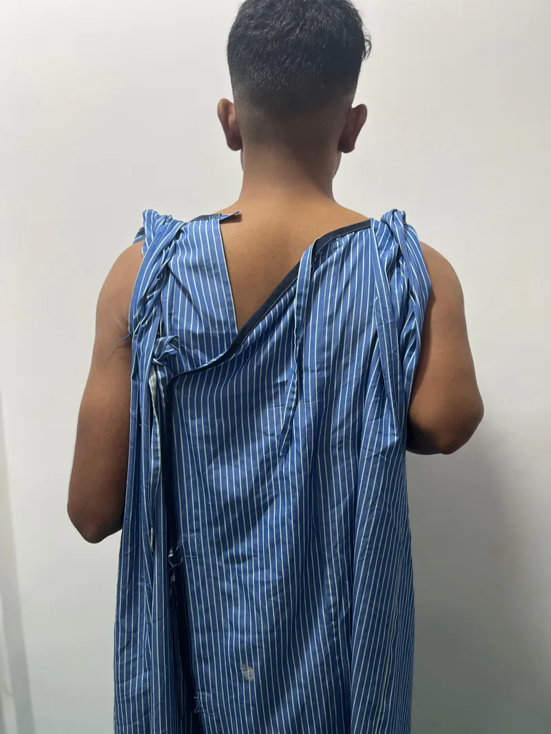
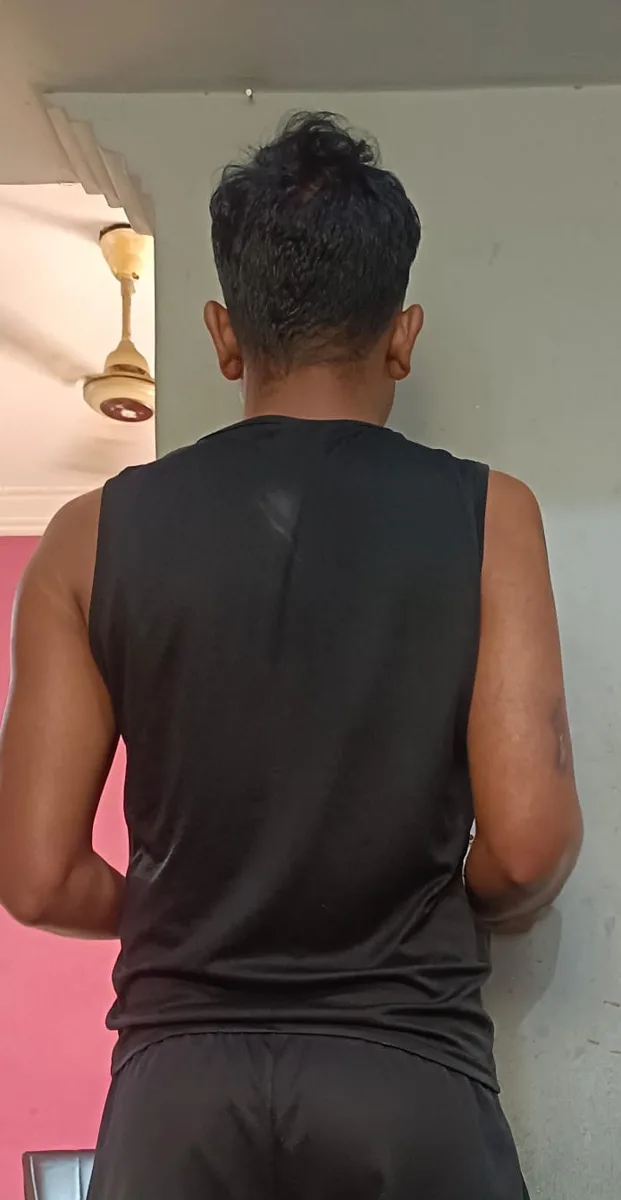
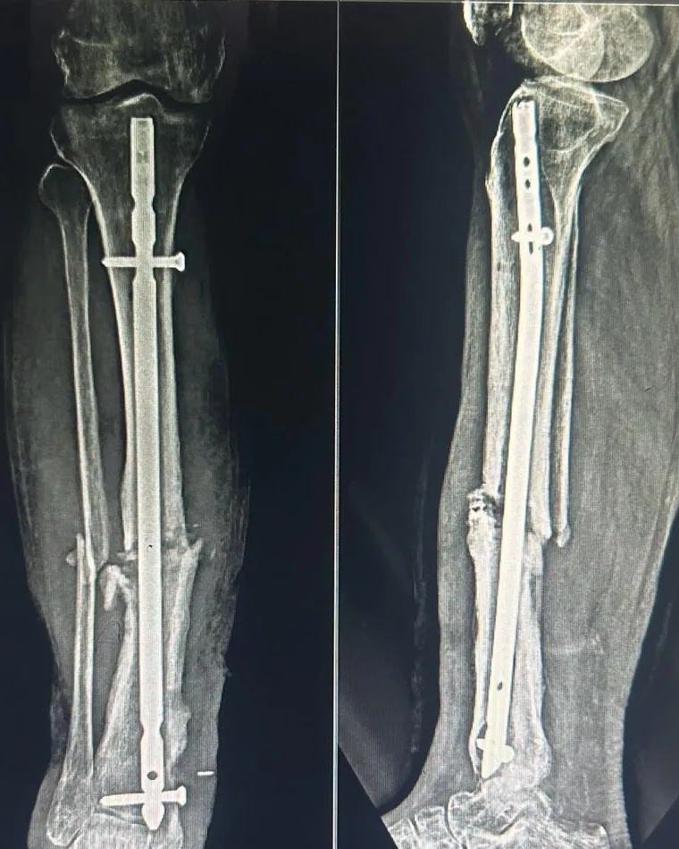
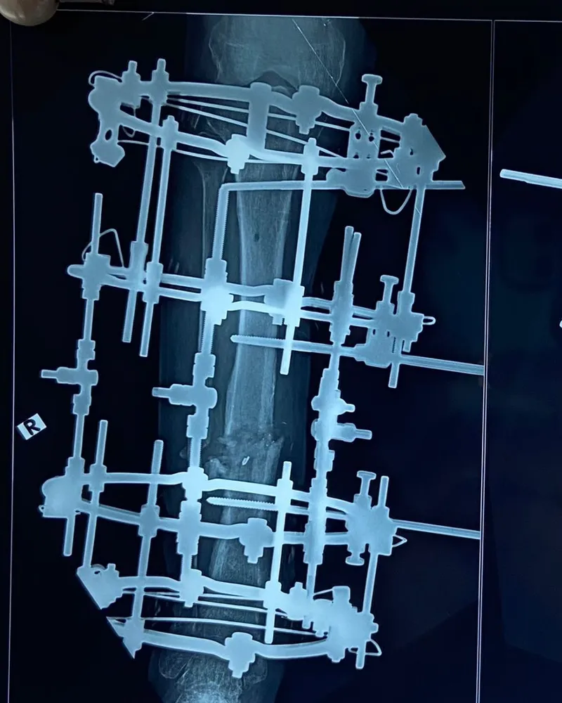
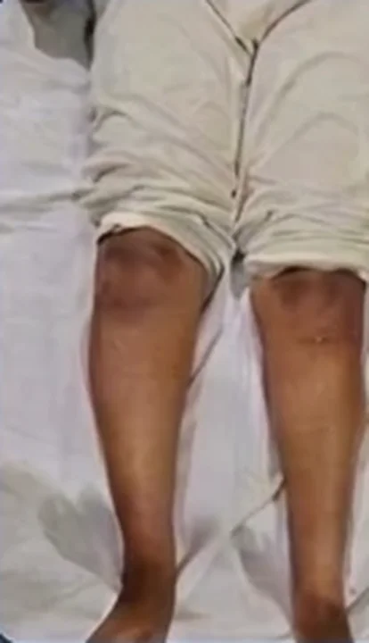
Presentation: knee pain with a visible bow-legged (varus) deformity that limited walking and daily activity.
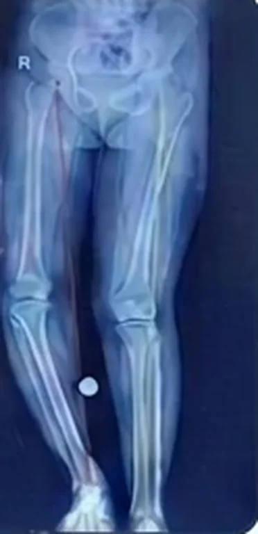
Findings: a full-length standing X-ray showed severe varus deformity with the mechanical axis (the weight-bearing line from hip to ankle) shifted to the inner side of the knee, overloading that compartment. Measurement showed the deformity came from both the thigh bone (femur) and the shin bone (tibia).
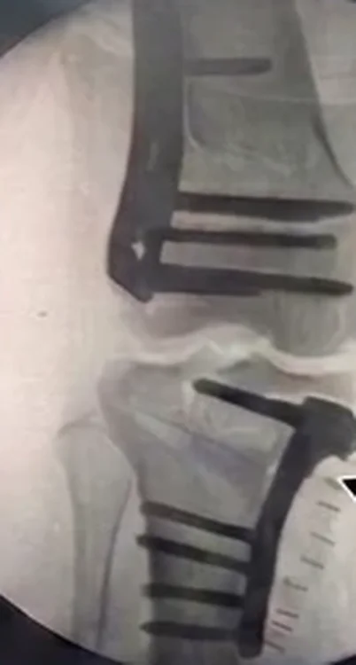
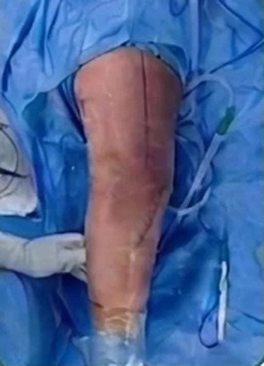
Procedure: osteotomy at both levels (double-level osteotomy): controlled bone cuts in the lower femur and the upper tibia to correct the deformity where it actually lies, each fixed with a plate. Correcting at both levels keeps the knee joint line level, rather than over-correcting at one bone.
Recovery: early knee movement, protected weight-bearing until the osteotomies healed, then progressive strengthening.
Why it matters: correcting alignment unloads the worn side of the knee and can delay the need for knee replacement in suitable patients. Read more about double-level osteotomy, high tibial osteotomy and bow legs and knock knees.
For patient education only. Illustrative problems are shared with written patient consent, with identifying details removed. Every patient is different and individual outcomes vary.


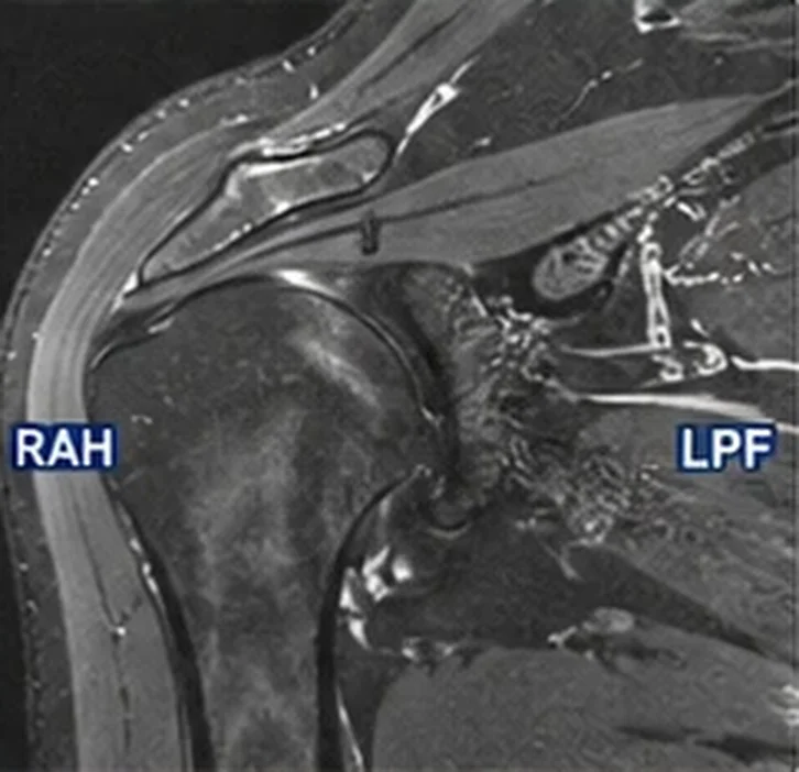 Problem
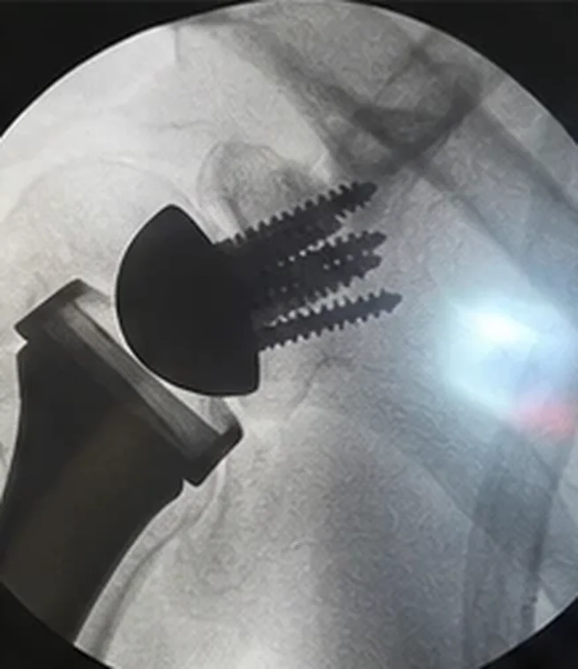 Solution