


 By Dr. Himanshu Singh, MBBS, D'Ortho, DNB (Orthopaedics)
The meniscus is a C-shaped cushion between the thigh bone and shin bone. Each end of the meniscus — its root — is anchored firmly to the bone. These roots let the meniscus act like a hoop: when you stand, it stretches outward and spreads your body weight over a wide area of cartilage.
A meniscus root tear detaches that anchor. The most common type is the medial meniscus posterior root tear (MMPRT), at the back of the inner side of the knee. It is common in middle-aged adults, women, and people with bow legs, and in daily activities that involve deep squatting or sitting cross-legged on the floor.
The classic story: a "pop" while squatting
- A sudden painful pop at the back of the knee while squatting, sitting cross-legged, getting up from the floor or on stairs
- Pain on the inner side of the knee when walking, especially on stairs
- Often only mild swelling — so it is frequently mistaken for a sprain
- In the weeks that follow, increasing pain on standing and walking
Why a root tear is a big deal
When the root tears, the hoop is broken. The meniscus can no longer hold load and is pushed out of the joint — called meniscal extrusion. In a cadaver study, a complete root tear raised peak pressure on the inner-side cartilage as much as removing the entire meniscus did.[1]
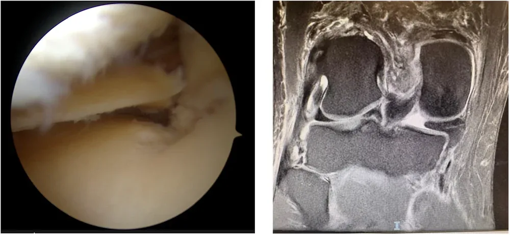
The cartilage underneath is suddenly overloaded. Over months, this can cause:
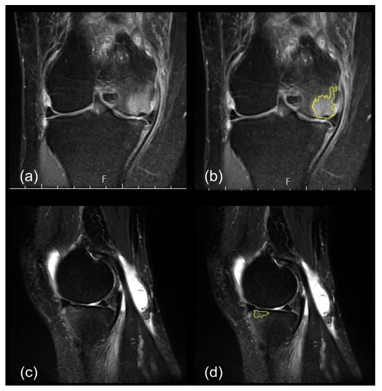
- Bone marrow oedema (bone bruising) under the cartilage
- Subchondral insufficiency fracture of the knee (SIFK) — a small collapse of the bone surface, often misread as "spontaneous osteonecrosis"
- Rapid cartilage loss and medial (inner-side) osteoarthritis
The bow-leg (varus) factor
In a varus (bow-legged) knee, the line of body weight passes through the inner side of the knee — the same side as the torn root. Varus alignment both increases the chance of a root tear and speeds up arthritis once the cushion is lost. It becomes a vicious cycle:
- Varus alignment overloads the inner compartment
- The overloaded meniscus root tears
- The meniscus extrudes and stops protecting the cartilage
- The cartilage wears, the inner joint space narrows, and the bow leg worsens
- Even more load goes through the inner side
Research on patients treated without surgery has found that older age and greater meniscal extrusion predict faster progression to arthritis.
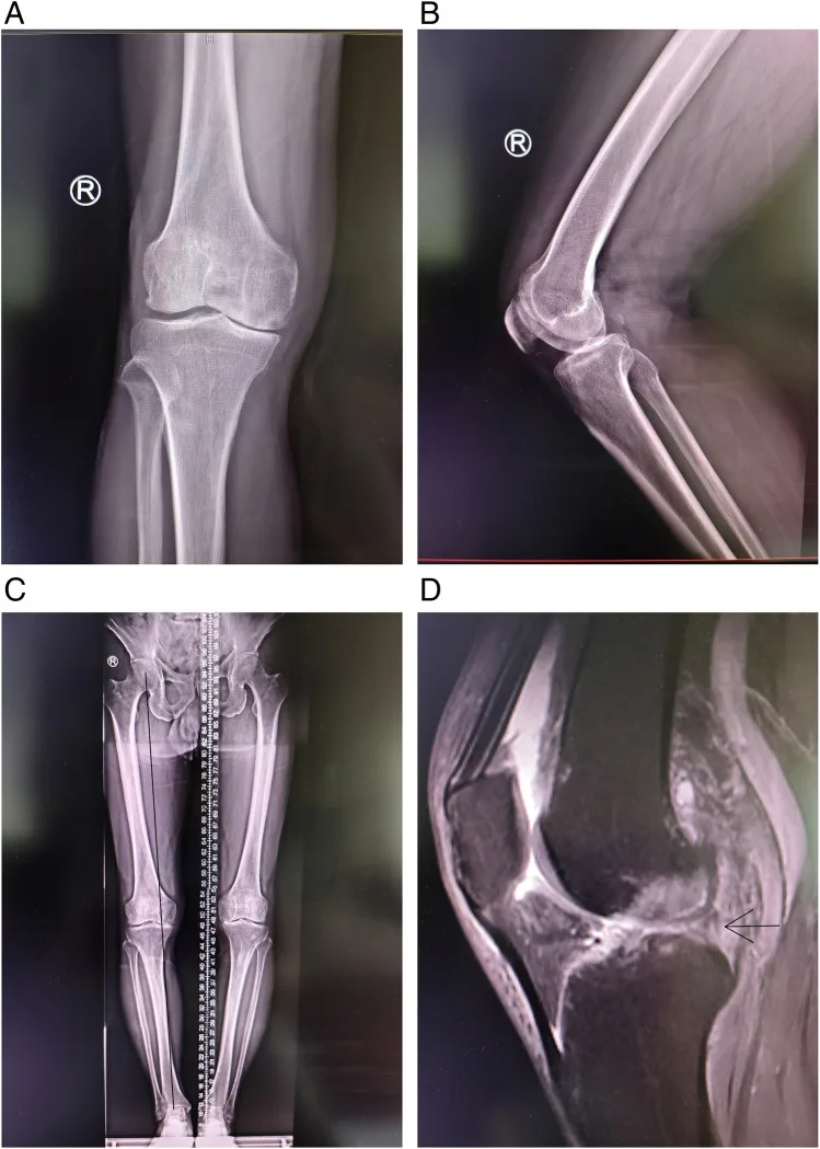
Diagnosis: the MRI signs
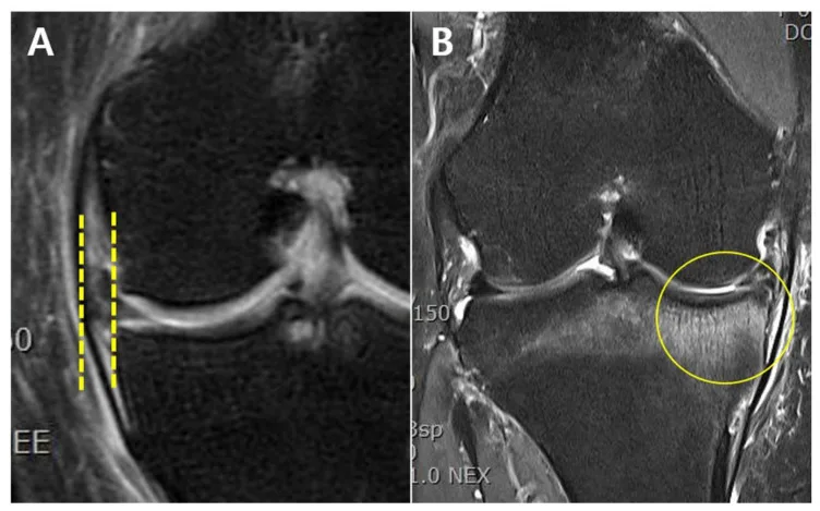
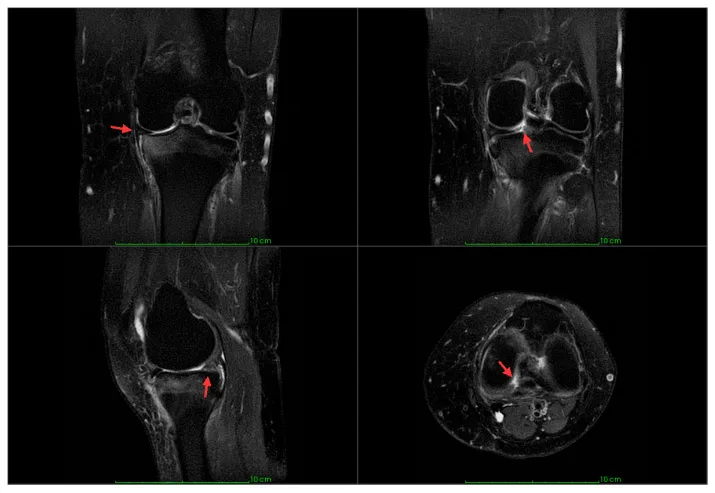
An MRI of the knee is the key test. Signs that point to a root tear include:
- Meniscal extrusion — the meniscus pushed out beyond the edge of the tibia (more than 3 mm is significant)
- Ghost sign — the posterior part of the meniscus "disappears" on sagittal images
- Cleft or radial tear at the root on coronal and axial images
- Bone marrow oedema in the medial tibia or femur
A full-length standing X-ray (hip to ankle) measures leg alignment, which determines whether a bow-leg correction is needed alongside the repair.
Treatment options
Root repair — the main goal is to restore the hoop
For an active patient with good cartilage, arthroscopic transtibial pull-out repair reattaches the root to its bony footprint: sutures are passed through the root and pulled through a small tunnel in the tibia, then fixed. A centralisation stitch may be added to pull an extruded meniscus back into place.
Root repair plus high tibial osteotomy (HTO)
When the knee is bow-legged, repairing the root alone leaves it under the same overload that caused the tear. A high tibial osteotomy at the same sitting shifts weight toward the healthy outer side, protecting the repair and the cartilage. For deformity in both the femur and tibia, a double-level osteotomy may be needed.
Non-surgical care
Activity modification, physiotherapy, weight management and pain relief are reasonable for patients with established arthritis or low demands. These ease symptoms but do not reattach the root.
When arthritis has already set in
If the cartilage is already badly worn, repair is less helpful. A partial knee replacement (UKR) or total knee replacement may then be the best way to relieve pain.
See a real case
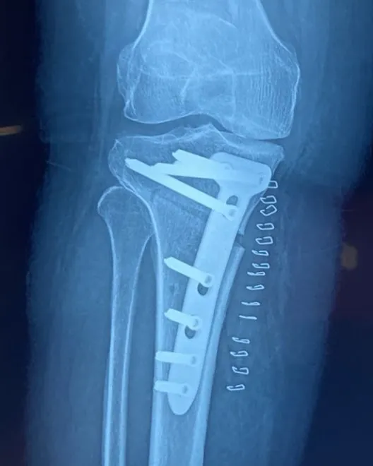
Shared with written patient consent, identifying details removed. Individual outcomes vary.
Recovery after root repair
Typical timeline in my practice; your plan may differ.
| Time | What to expect |
|---|---|
| 0–6 weeks | Brace; touch or partial weight bearing; bending limited to protect the repair |
| 6–8 weeks | Full weight bearing; range of movement restored |
| 3 months | Strengthening; cycling and swimming |
| 4–6 months | Return to most activities; avoid deep squatting early |
When an HTO is added, the bone healing timeline (about 6–8 weeks before full weight bearing) applies as well.
The message: don't ignore the pop
A painful pop at the back of the knee while squatting, followed by inner-knee pain, deserves an early specialist assessment and MRI. Early repair — with alignment correction when the leg is bowed — gives the best chance of saving your own knee.
References
- Allaire R, Muriuki M, Gilbertson L, Harner CD. Biomechanical consequences of a tear of the posterior root of the medial meniscus. Similar to total meniscectomy. J Bone Joint Surg Am. 2008;90(9):1922-31. PMID 18762653. pubmed.ncbi.nlm.nih.gov/18762653/
Frequently asked questions
What does a meniscus root tear feel like?
Many patients describe a sudden painful pop at the back of the knee while squatting, sitting cross-legged, getting up from the floor or climbing stairs, followed by pain on the inner side of the knee when walking. Swelling is often mild, so it is easily dismissed as a sprain.
Why is a root tear more serious than other meniscus tears?
The root anchors the meniscus to bone. When it tears, the meniscus can no longer absorb load — biomechanically it behaves as if the whole meniscus had been removed. The cartilage on the inner side of the knee is then overloaded and can wear out quickly.
Can a meniscus root tear heal on its own?
A complete root tear does not heal back to bone by itself. Physiotherapy and pain relief can ease symptoms, but studies show that, without repair, arthritis often progresses — particularly with older age, bow legs and meniscal extrusion on MRI.
Why does my bow leg (varus) matter?
In a varus knee, more body weight passes through the inner compartment. A root tear removes the meniscus cushion from exactly that side. The two together speed up cartilage loss, so correcting alignment with a high tibial osteotomy is often combined with root repair.
Is there a time limit for root repair?
Results are better when the repair is done early, before cartilage damage and meniscal extrusion become established. Once advanced arthritis has developed, partial or total knee replacement may be the better option.
Medical disclaimer: this article is general information, not a diagnosis. Every injury is different — please consult an orthopaedic surgeon with your scans before deciding on treatment.