


 By Dr. Himanshu Singh, MBBS, D'Ortho, DNB (Orthopaedics)
A kneecap (patellar) dislocation happens when the kneecap slips out of its groove, almost always to the outer side, typically on twisting or landing. It is most common in teenagers and young adults. The knee swells, and the kneecap may pop back by itself or need to be put back.
After the first dislocation, many people go on to have repeat dislocations or a constant feeling that the kneecap is "about to go". Repeated episodes damage the cartilage behind the kneecap. The aim of treatment is to find why the kneecap is unstable and fix the cause.
Why does the kneecap dislocate?
The main anatomical risk factors are:
- Trochlear dysplasia: a shallow or flat groove at the end of the femur. This is the most important risk factor.
- Patella alta: a kneecap that sits too high, so it engages the groove late
- Increased TT–TG distance: the tendon's attachment on the shin lies too far outwards, pulling the kneecap out
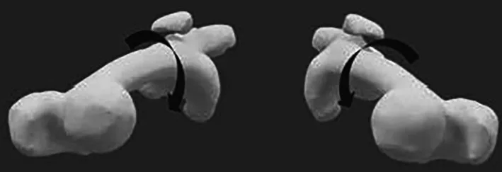
- Excessive femoral anteversion: an inward twist of the thigh bone
- Knock-knee (valgus) alignment
- Torn MPFL: the medial patellofemoral ligament, the main "seat-belt" holding the kneecap in, tears in almost every dislocation
Diagnosis
- X-rays: kneecap height, groove shape, any bony fragments
- MRI: MPFL tear, cartilage damage, loose fragments, and groove shape
- CT scan: TT–TG distance and the twist (rotation) of the femur and tibia when surgery is being planned
- Full-length standing X-ray if a knock-knee is suspected
Treatment
First dislocation, no fragment: usually a brace, then physiotherapy for the quadriceps and hip muscles, and a gradual return to sport.
Loose cartilage or bone fragment: early arthroscopy to fix or remove the fragment, often with ligament repair or reconstruction.
Recurrent dislocation: surgery, tailored to the risk factors found:
| Problem found | Usual procedure |
|---|---|
| Torn MPFL, mild or borderline risk factors | MPFL reconstruction alone |
| Tendon attachment too far out, or kneecap too high | Add tibial tubercle osteotomy (moving the attachment point) |
| Marked inward twist of the femur | Add derotation osteotomy of the femur |
| Knock-knee alignment | Add distal femoral osteotomy |
| Severe trochlear dysplasia | Trochleoplasty (deepening the groove) in selected cases |
A 2025 review concluded that combined procedures should be reserved for carefully selected patients with substantial anatomical risk factors, while isolated MPFL reconstruction may be adequate when the risk factors are milder.
MPFL reconstruction: what's involved
- Keyhole examination of the knee to assess and treat cartilage damage
- A hamstring tendon graft is taken through a small incision
- The graft is fixed to the kneecap and to a precise point on the femur, checked with X-ray guidance. Exact placement is what makes the reconstruction work without over-tightening
- The graft is tensioned with the knee bent so that the kneecap tracks centrally
Recovery after MPFL reconstruction
Typical timeline in my practice; your plan may differ.
| Time | What to expect |
|---|---|
| 0–2 weeks | Brace; walking with crutches; early knee bending as advised |
| 2–6 weeks | Weight-bearing as comfortable; bend to 90° and beyond |
| 6–12 weeks | Off crutches; strengthening and balance work |
| 3–4 months | Running in a straight line |
| 5–6 months | Return to sport after strength and hop testing |
Recovery is longer when an osteotomy is added, because the bone needs to heal first.
Results
In a 2026 study of isolated MPFL reconstruction for recurrent instability, repeat dislocation rates were low (4–7%) and function scores improved substantially, even in patients with borderline anatomical risk factors. Most patients return to their previous sport.
Why alignment and rotation matter
Kneecap instability is often a problem of the whole leg, not just the knee. Training in limb reconstruction and deformity correction means rotation, alignment and kneecap height are measured and corrected where needed, rather than relying on a ligament graft that is under constant strain.
Frequently asked questions
Does a first kneecap dislocation need surgery?
Usually not. Most first-time dislocations are treated with a brace or support, then physiotherapy to strengthen the thigh and hip muscles. Early surgery is considered when a piece of cartilage or bone has broken off inside the knee, or when scans show major anatomical risk factors that make repeat dislocation very likely.
What is MPFL reconstruction?
The medial patellofemoral ligament (MPFL) is the main restraint that stops the kneecap sliding outwards, and it tears in almost every dislocation. MPFL reconstruction replaces it with a tendon graft, usually from the hamstrings, fixed to the kneecap and the femur at the precise anatomical points.
When is more than MPFL reconstruction needed?
If the groove for the kneecap is very shallow (severe trochlear dysplasia), the kneecap sits very high, the shin's attachment point is far to the outside, the thigh bone is twisted inwards or the leg is knock-kneed, those problems may need correcting too, with a tibial tubercle osteotomy, derotation osteotomy or distal femoral osteotomy. For milder risk factors, MPFL reconstruction alone usually works well.
Medical disclaimer: this article is general information, not a diagnosis. Every injury is different — please consult an orthopaedic surgeon with your scans before deciding on treatment.