

 By Dr. Himanshu Singh, MBBS, D'Ortho, DNB (Orthopaedics)
In a knock-knee (genu valgum) the knees come together while the ankles stay apart. The weight-bearing line passes through the outer (lateral) side of the knee instead of the centre. Over time this overload can cause outer-knee pain, early lateral arthritis, and it can pull the kneecap outwards, contributing to kneecap dislocation.
In most adults the deformity comes from the lower end of the thigh bone (distal femur). A distal femoral osteotomy (DFO) corrects it at that level, just as HTO corrects bow legs at the tibia. It is joint preservation: you keep your own knee.
Who benefits?
- Outer-knee pain or early lateral compartment arthritis with valgus alignment
- Kneecap instability where valgus alignment contributes, often combined with ligament reconstruction
- Overload after removal of the outer meniscus, or to protect a meniscus or cartilage repair
- Young, active adults, typically under about 55–60, with a healthy inner compartment and good knee movement
- Knock-knees from childhood that were never corrected (see knock-knee and bow-leg correction)
Finding where the deformity is
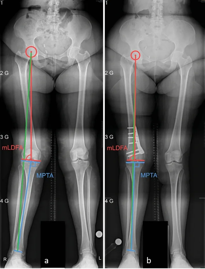
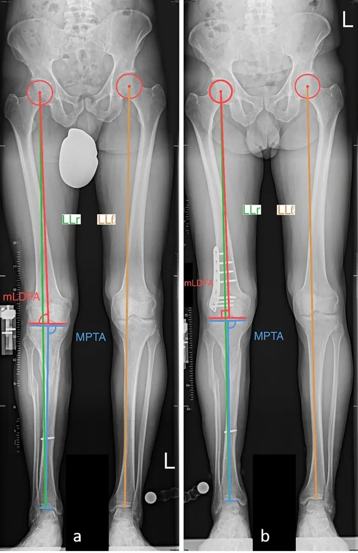
The most important step is to measure where the knock-knee actually comes from. On a full-length standing X-ray (hip to ankle):
- The mechanical axis shows how far the weight-bearing line has moved outwards
- The femoral joint angle (mLDFA) shows whether the femur is the cause (a low angle, often around 84° or less)
- The tibial joint angle (MPTA) shows whether the tibia also contributes
If the deformity is in the femur, DFO is the right operation. If both the femur and the tibia contribute, correcting only one bone can tilt the knee joint line, so a double-level osteotomy may be better. MRI checks the cartilage, menisci and ligaments, and a CT scan is added when rotation of the thigh bone also needs correcting.
The two main techniques
| Medial closing-wedge DFO | Lateral opening-wedge DFO | |
|---|---|---|
| How | A small wedge of bone is removed from the inner femur and closed | The bone is cut from the outer side and opened to the planned angle |
| Healing | Bone-on-bone contact: tends to heal faster | Gap fills with new bone; graft sometimes added |
| Leg length | Shortens slightly | Lengthens slightly: useful if that leg is already short |
| Typical use | Most valgus corrections | Smaller corrections, or when length is needed |
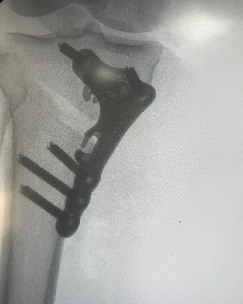
Both are fixed with a locking plate. The correction is calculated so that the weight-bearing line returns to the centre of the knee. In arthritis it is not over-corrected into bow legs.
Risks to know about
DFO is effective but it is real bone surgery. In one series of 28 DFOs, minor complications occurred in 25% and major complications in 14%. Delayed bone healing occurred in 18% and non-union in 4%, and some patients later needed plate removal. Careful planning, stable fixation, stopping smoking and following weight-bearing advice all reduce these risks.
See a real case
Deformity (alignment) correction with limb lengthening
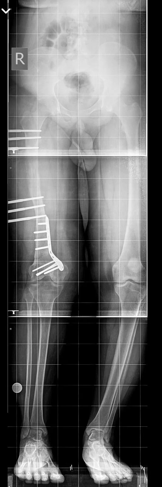
Knock knees with a short leg: alignment correction and lengthening
A young adult with knock knees and a shorter leg, causing knee pain and an uneven walk. Both the alignment and the length were corrected, planned on full-length standing X-rays.
Arthroscopic meniscus repair + realignment osteotomy HTO
Knee pain in a varus knee: meniscus repair with realignment High Tibial Osteotomy (HTO)
A woman in her late fifties with right knee pain, a meniscus tear and bow legs alignment. The joint was preserved by repairing the meniscus and correcting the alignment.
Shared with written patient consent, identifying details removed. Individual outcomes vary.
Recovery
Typical timeline in my practice; your plan may differ.
| Time | What to expect |
|---|---|
| Day 1 | Walking with crutches; knee bending exercises start |
| 0–6 weeks | Protected (partial) weight-bearing as advised; quadriceps and movement physiotherapy |
| 6–8 weeks | X-ray check; progress to full weight-bearing |
| 3 months | Most daily activities, cycling, swimming |
| 6 months | Running and sport once healing is confirmed |
Results
Well-selected patients get relief of outer-knee pain and a straighter leg. A 2026 study of young patients having isolated DFO reported high rates of return to sport. In arthritis, DFO is designed to delay knee replacement, and replacement can still be done later if needed.
Why deformity training matters
Osteotomy is deformity surgery. Training in limb reconstruction means the whole leg is analysed: alignment, rotation and leg length. The correction is then made at the right bone, to the right amount, and the knee joint line is kept level.
Frequently asked questions
Who needs a distal femoral osteotomy?
DFO is considered for knock-knee (valgus) alignment that comes from the lower femur and causes outer-knee pain or early arthritis, kneecap dislocation, or overload after an outer meniscus has been removed. It is also used to protect cartilage or meniscus repairs in a valgus knee. Patients are usually younger and active, with the inner side of the knee still healthy.
What is the difference between a closing-wedge and an opening-wedge DFO?
In a medial closing-wedge DFO a small wedge of bone is removed from the inner side of the femur; it heals quickly and slightly shortens the leg. In a lateral opening-wedge DFO the bone is opened from the outer side; it slightly lengthens the leg, which can help when that leg is already shorter. The choice depends on the amount of correction, leg lengths and the knee's other problems.
How long does it take to recover from DFO?
Most patients use crutches for about 6 weeks, return to desk work in 4–6 weeks, and to impact sport at around 6 months once X-rays confirm healing. Studies of young patients show high rates of return to sport after DFO.
Medical disclaimer: this article is general information, not a diagnosis. Every injury is different — please consult an orthopaedic surgeon with your scans before deciding on treatment.