

 By Dr. Himanshu Singh, MBBS, D'Ortho, DNB (Orthopaedics)
Most bow-leg (varus) deformities in adults come from the shin bone (tibia), and a high tibial osteotomy corrects them well. But in a significant number of patients, especially with severe bow legs, the thigh bone (femur) is also bent. For them, correcting the whole deformity in the tibia alone causes a new problem: a tilted knee joint.
A double-level osteotomy (DLO) corrects both bones, the distal femur and the proximal tibia, so each is straightened where the deformity lies.
The problem of joint-line obliquity
The knee works best when its joint surface is roughly horizontal when you stand. When a large correction is made in the tibia alone to compensate for a femoral bow:
- The joint line becomes oblique (tilted)
- The femur tends to slide on the tibia, creating shear forces on the cartilage
- The results of the osteotomy may be less durable
A DLO avoids this by sharing the correction between the two bones.
Who needs a DLO?
- Severe varus (bow-leg) deformity
- Deformity analysis shows both the femur and tibia contribute (abnormal femoral and tibial joint angles, such as the LDFA and MPTA)
- Correcting the tibia alone would leave the knee joint line tilted beyond an acceptable limit
- Medial knee arthritis or cartilage damage in an active patient who wants to keep their own knee
- Some patients with post-traumatic or developmental deformities (for example, after childhood rickets or Blount's disease)
Planning: the most important step
Double-level correction is deformity surgery, rooted in the principles of limb reconstruction:
- Full-length standing X-rays of both legs
- Measurement of the mechanical axis and the joint orientation angles of the femur and tibia
- Identifying the centre of rotation of angulation (CORA) in each bone
- Digital planning of how much correction goes into each bone, aiming for a straight mechanical axis and a level joint line
- MRI of the knee for cartilage and meniscus, and sometimes a CT to assess rotation
The operation
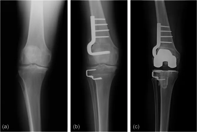
- Distal femoral osteotomy (DFO): a precise cut in the lower femur, fixed with a locking plate
- High tibial osteotomy (HTO): usually a medial opening wedge, fixed with a locking plate
- Both are generally done in one sitting, with arthroscopy to treat the meniscus or cartilage if needed
- Intra-operative X-ray guidance confirms the alignment before fixation
In selected cases, especially with rotational deformity or very large corrections, gradual correction with an external (hexapod) fixator may be preferred.
See a real case
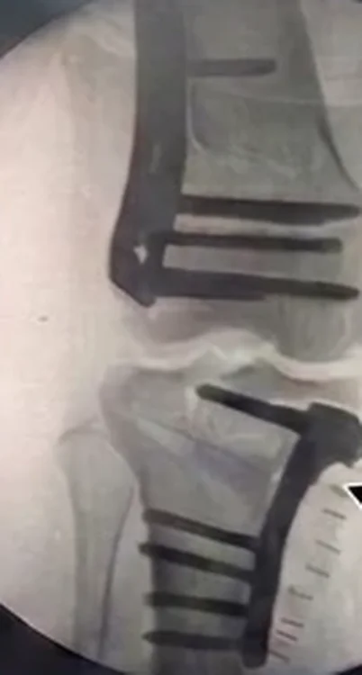
Shared with written patient consent, identifying details removed. Individual outcomes vary.
Recovery
Typical timeline in my practice; your plan may differ.
| Time | What to expect |
|---|---|
| Day 1 | Walking with crutches; knee movement exercises |
| 0–6 weeks | Protected weight bearing; physiotherapy for movement and strength |
| 6–10 weeks | X-ray checks; progress to full weight bearing |
| 3 months | Walking normally; cycling and swimming |
| 4–6 months | Return to sport once both osteotomies have healed |
Plates may be removed after a year if they cause irritation.
The benefit
DLO lets active patients with complex bow-leg deformity keep their own knee, with a straight leg and a level joint. It relieves pain and protects the cartilage, and keeps every future option, including knee replacement, open.
Related osteotomies
- High tibial osteotomy (HTO): when the bowing comes mainly from the tibia
- TCVO: advanced inner-knee arthritis with a widened, unstable outer joint
- Distal femoral osteotomy (DFO): knock-knee deformity coming from the femur
Frequently asked questions
What is a double-level osteotomy?
It is a realignment operation where both the lower femur (distal femoral osteotomy) and the upper tibia (high tibial osteotomy) are corrected, usually in one sitting, so that each bone is straightened where its deformity actually lies.
Why not just do a high tibial osteotomy?
If a large part of the bow leg comes from the femur, correcting everything in the tibia over-tilts the knee joint line. An oblique joint line causes abnormal shear forces on the cartilage and may give poorer results. Correcting both bones keeps the joint line level.
Is recovery longer than a single osteotomy?
Recovery is similar to or slightly longer than a single osteotomy. Most patients walk with crutches from the first day, progress to full weight bearing over 6–10 weeks, and return to sport after both bones have healed, usually 4–6 months.
Medical disclaimer: this article is general information, not a diagnosis. Every injury is different — please consult an orthopaedic surgeon with your scans before deciding on treatment.