


 By Dr. Himanshu Singh, MBBS, D'Ortho, DNB (Orthopaedics)
Bow-leg (varus) knee arthritis wears away the inner (medial) side of the knee first. In the early stages a high tibial osteotomy (HTO) can shift weight to the healthy outer side. In more advanced knees, though, something else happens: as the inner side collapses, the outer side of the joint opens up like a hinge, the ligaments slacken and the knee wobbles from side to side. A standard HTO can straighten the leg, yet still leave that outer joint gaping.
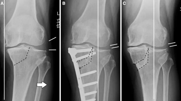
The tibial condylar valgus osteotomy (TCVO) was developed in Japan in 1989 for exactly this problem. It corrects the bow leg and closes the gap inside the joint, so the knee is aligned and stable again, while you keep your own knee.
Who is TCVO for?
TCVO is considered for knees with:
- Medial (inner) compartment arthritis, often moderate to advanced (Kellgren–Lawrence grade III–IV)
- Bow-leg alignment with the weight-bearing line passing through the inner side of the knee
- A widened, subluxated outer joint on X-ray, and side-to-side (varus–valgus) looseness
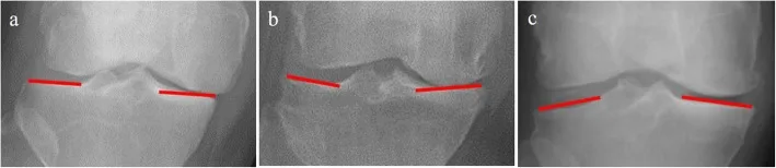
- A convex "pagoda-shaped" tibial plateau, with the joint lines converging by more than about 5° on standing X-ray
- Reasonable knee bending: at least about 90°
- Healthy cartilage on the outer side, because that is where the weight will be moved
It is particularly useful for patients who have been told they need a knee replacement but are active, prefer to keep their own joint, or are not yet ready for replacement.
How is it different from HTO?
| High tibial osteotomy (HTO) | Tibial condylar valgus osteotomy (TCVO) | |
|---|---|---|
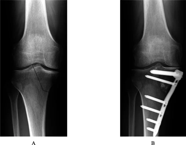
| Where the cut is | Across the tibia, below the joint | L-shaped, running up into the centre of the joint (between the tibial spines) |
| What it corrects | Leg alignment | Leg alignment plus joint congruity and stability |
| Best for | Earlier arthritis, stable knee | Advanced arthritis, widened outer joint, unstable knee |
| Typical patient | Younger, active | Often older, with more advanced disease |
In a 2024 comparison, patients having TCVO were older and had more advanced arthritis, more bow-leg deformity and more joint instability than those having HTO. Even so, alignment and stability improved to comparable levels in both groups, and patient-reported scores recovered well in both by one year.
Planning: the key to a good result
- Full-length standing X-rays (hip to ankle) to measure the mechanical axis and decide the correction angle
- Stress X-rays (varus and valgus) to measure how much the joint opens and how unstable it is
- Assessment of the tibial plateau shape and the joint-line convergence angle
- MRI when needed, to check the outer compartment cartilage, meniscus and ligaments
- Calculation of the correction so that the weight-bearing line moves just past the centre of the knee, to around 60% of the joint width
If the required correction is larger than TCVO alone can provide, it can be combined with an additional osteotomy (for example an opening-wedge HTO) at the same operation, an approach described by the groups that developed the technique.
The operation
- Under X-ray guidance, an L-shaped cut is made from the inner side of the upper tibia, turning upwards to end at the centre of the knee joint, between the tibial spines
- The cut is gradually opened with a spreader until the outer tibial joint surface meets the outer femur again: the gap closes and the knee becomes stable in extension
- The correction is held with a locking plate. A circular (Ilizarov-type) external fixator is an alternative in selected cases
- The opened space is filled with bone graft or a bone-graft substitute
- Knee arthroscopy can be done at the same sitting to deal with loose bodies or meniscus tears
Because part of the cut enters the joint, TCVO needs precise X-ray-guided technique, the same skills used in limb reconstruction and deformity correction.
Recovery
Typical timeline in my practice; your plan may differ.
| Time | What to expect |
|---|---|
| Day 1–2 | Standing and walking with a walker or crutches; knee bending exercises begin |
| 0–6 weeks | Weight-bearing increased step by step as advised; quadriceps and movement physiotherapy |
| 6–8 weeks | X-ray check of healing; most patients walk without crutches |
| 3 months | Daily activities, longer walks, stationary cycling |
| 6–12 months | Continued improvement as the bone remodels and strength returns |
Results
In a 5-year study, average pain scores fell from 73 to 13 (out of 100) and WOMAC arthritis scores improved from 52 to 14. The weight-bearing line moved from the inner edge of the knee to about 60% across, and the joint-line convergence fell from 6° to 1°, meaning the outer joint had closed and the knee was more stable. Knee bending tends to stay a little lower after TCVO than after HTO, so good pre-operative movement matters.
Individual results vary. TCVO does not stop all future wear, but it can relieve pain and delay knee replacement, and a knee replacement can still be done later if needed.
HTO, TCVO, DLO or knee replacement?
- HTO: earlier medial arthritis, stable knee, bowing in the tibia
- TCVO: advanced medial arthritis with a widened outer joint and instability
- Double-level osteotomy (DLO): severe bowing coming from both the femur and the tibia
- Partial or total knee replacement: arthritis in more than one compartment, stiff knees, or when osteotomy is not suitable
The right choice depends on your X-rays, how your knee moves and what you want to do. A careful assessment on full-length X-rays is the first step.
Frequently asked questions
What is the difference between TCVO and HTO?
A standard high tibial osteotomy (HTO) cuts the tibia below the joint and tilts the whole knee. TCVO uses an L-shaped cut that runs up into the centre of the knee joint, so it corrects the bow leg and also closes the widened, unstable outer side of the joint. TCVO suits more advanced arthritis with joint instability; HTO suits earlier arthritis with a stable knee.
Am I too old for TCVO?
TCVO has been used successfully in patients in their sixties and seventies with advanced medial arthritis. What matters more than age is the shape of the joint, good knee bending (at least about 90°), healthy outer compartment cartilage and general fitness. Each knee is assessed on full-length X-rays and stress X-rays.
How long is recovery after TCVO?
Patients walk with support from the first days, increase weight-bearing over about 6 weeks as the X-rays allow, and most return to daily activities in 3 months. Full benefit, as the bone heals and the muscles recover, usually takes 6 to 12 months.
Medical disclaimer: this article is general information, not a diagnosis. Every injury is different — please consult an orthopaedic surgeon with your scans before deciding on treatment.